Littermate

Reference

The Library

Eight peptides studied in rodents and in cell culture. Each profile says what the compound is, what the animal literature reports, and where the evidence runs out.

Last reviewed 3 August 2026. Edited by Nadia Bergeron, fact-checked by Danny Whitcombe.

Tissue Repair Immunomodulation Dermal Metabolic Host Defense Neuro

01   BPC-157  /  Synthetic pentadecapeptide  /  Rat (Sprague-Dawley, Wistar)

BPC-157

A fifteen-residue sequence with a large rodent literature and almost no independent one.

What it is

BPC-157 is a synthetic chain of fifteen amino acids. Its published origin story is that the sequence corresponds to a partial region of a protein described in human gastric juice, which is where the name comes from: body protection compound. That parent protein has never become a well-characterised entity in the wider biochemical literature, so it is more accurate to describe BPC-157 as a synthetic sequence with a proposed gastric origin than as a fragment of a known human protein.

The property most often claimed for it is stability in acidic conditions, which is the basis for the oral administration used in much of the rodent work. It has no established receptor. Proposed mechanisms in the published papers are pathway-level and varied: nitric oxide signalling, vascular endothelial growth factor expression, and effects on early vessel formation around an injury.

What the animal literature reports

The bulk of the literature is rat surgery. The recurring musculoskeletal models are Achilles tendon transection in Sprague-Dawley and Wistar rats, medial collateral ligament transection, and quadriceps muscle crush injury. Reported outcomes are histological healing scores read from stained sections and biomechanical load-to-failure testing of the repaired tissue, with treated animals reported to score better on both.

The gastrointestinal work is older and larger in volume: ethanol-induced and stress-induced gastric lesions in rats, chemically induced colitis, and surgically created fistula models, with reported reductions in lesion area and faster closure. There is a further body of rat work on vascular anastomosis and on injury to the blood supply itself.

Cell culture work is thinner and mostly supportive: cultured rat and human tendon fibroblasts reported to show increased migration and F-actin formation on exposure. Mouse work exists but is a small minority of the corpus.

Where the evidence thins

The dominant problem is not any single paper. It is that a very large share of this literature comes from one research network, centred on a Zagreb group and its collaborators, publishing across the 1990s, 2000s and 2010s. Papers from a single network are one body of work. Counting them as dozens of confirmations is a category error, and it is the error most often made about this compound.

Within the papers, group sizes are usually small, blinding of histological scoring is frequently not described, and the outcome measures vary enough between studies that pooling them is not meaningful. Independent replication by unaffiliated laboratories is sparse, and where third parties have looked at specific claims the picture has been less clean than the original reports.

There are no completed, published, randomized controlled human trials establishing efficacy for any indication, and no established human safety dataset. The honest summary is that the human evidence base is essentially absent, and that everything interesting about this compound is currently a rodent finding awaiting an independent laboratory.

Evidence snapshot: BPC-157

Model organisms Rat, predominantly; some mouse; cultured tendon fibroblasts
Study scale Small. Group sizes typically single digits to low teens
Human trial status No completed, published randomized controlled trials
Replication status Concentrated in one research network; independent replication sparse

Further reading on this page: One lab, many tendons: the replication problem under the BPC-157 literature
02   Thymosin β4  /  Actin-sequestering peptide  /  Mouse (wound, cardiac); rat

Thymosin β4, and the fragment called TB-500

A real and abundant intracellular protein, and a short fragment that secondary sources treat as the same thing.

What it is

Thymosin β4 is a naturally occurring peptide of forty-three residues, present at high concentration inside many cell types. Its established biochemical role is to bind monomeric actin and hold it in reserve, regulating how readily the cell builds actin filaments. That is a well-described function and is not in dispute.

TB-500 is a name attached to a short fragment corresponding to an actin-binding region of the parent peptide. The distinction matters and is routinely collapsed outside the primary literature. A result obtained with the full forty-three-residue peptide is not a result about a seven-residue fragment, and papers that study one generally do not study the other.

What the animal literature reports

Murine full-thickness dermal wound models are the most repeated setting, with reports of faster re-epithelialisation and higher vessel density in treated animals. Corneal injury models in rat and mouse report similar directional findings on epithelial closure.

The cardiac work is the most cited and the most contested. Mouse myocardial infarction models have reported activation of epicardium-derived cells, increased vessel formation in the border zone, and in some papers improvements in ejection fraction measured by echocardiography. There is also rodent work in neurological injury and in dry eye.

Cell culture supports the migration story: endothelial cells and keratinocytes are reported to migrate faster in its presence, which is what an actin-regulating peptide would be expected to do.

Where the evidence thins

Most reported effects are surrogate: vessel counts, marker expression, area measurements on a stained section. Those are not the same as durable functional recovery, and papers in this corner often report whichever of several plausible endpoints moved.

The cardiac progenitor findings were striking enough to attract direct scrutiny, and subsequent work in other laboratories did not consistently support the strongest version of the claim. That disagreement is in the published record and is a better guide to the state of the evidence than any single dramatic paper.

Human clinical work exists for the full-length peptide in narrow settings, principally ophthalmic surface disease and some wound contexts. It has been small and mixed. None of it is evidence about the fragment sold under a different name, and no randomized human trial of that fragment has been published.

Evidence snapshot: Thymosin β4

Model organisms Mouse (dermal wound, myocardial infarction); rat cornea; cultured endothelium
Study scale Small to moderate; larger in the cardiac literature
Human trial status Limited early-phase work on the full peptide only; mixed results
Replication status Wound findings broadly repeated; cardiac findings openly contested

Further reading on this page: What the murine infarct models measured when they measured thymosin beta-4

03   GHK-Cu  /  Copper-binding tripeptide  /  Rat and mouse dermal; cultured human fibroblasts

GHK-Cu

A genuine human plasma tripeptide whose reputation rests mostly on what it does in a dish.

What it is

GHK is glycyl-L-histidyl-L-lysine, a tripeptide first isolated from human plasma. It binds copper with high affinity, and the copper complex, written GHK-Cu, is the entity almost all of the research concerns. Unlike several other compounds in this Library, there is no ambiguity about whether the molecule occurs naturally in people. It does.

The proposed mechanism is partly delivery: the peptide carries copper, and copper is a required cofactor for enzymes involved in collagen and elastin cross-linking. The peptide is also reported to have signalling effects of its own on gene expression in cultured cells.

What the animal literature reports

The animal literature is dermal and mostly old. Rat and mouse full-thickness wound models from the 1980s and 1990s report faster closure and increased collagen deposition in treated wounds. There is also work in rabbit ear and pig skin, the latter being the more relevant model for human dermis in structural terms.

The much larger body of work is cell culture in cultured human dermal fibroblasts, where exposure is reported to raise type I collagen, elastin and glycosaminoglycan synthesis and to shift the balance of matrix metalloproteinases and their tissue inhibitors. Gene expression profiling in cultured cells has produced long lists of transcripts reported to move, which is where a great deal of the popular writing about this molecule originates.

Where the evidence thins

A cultured fibroblast sits in a dish with a known concentration applied directly to it. Dermis is a layered tissue behind a barrier that the molecule has to cross at all, in an amount nobody in the cell culture papers measured. The gap between those two situations is the whole question, and the rodent studies that would bridge it are small, dated, and generally silent on randomization and blinded assessment.

The copper problem is rarely addressed head-on. Copper delivery alone has effects on wound tissue. Very few studies include the control that would separate a peptide-specific effect from a copper-delivery effect, so most reported findings are compatible with either explanation.

Human evidence is mostly small cosmetic studies with subjective or photographic endpoints, frequently run or funded by manufacturers of the products tested. That is not a substitute for controlled trial evidence, and we do not treat it as one.

Evidence snapshot: GHK-Cu

Model organisms Rat and mouse dermal wounds; rabbit ear; pig skin; cultured human fibroblasts
Study scale Small animal studies; extensive but shallow cell culture
Human trial status Small cosmetic studies, largely industry-run, subjective endpoints
Replication status Cell culture findings widely repeated; animal work rarely repeated

Further reading on this page: Copper peptides and the distance between a fibroblast dish and skin

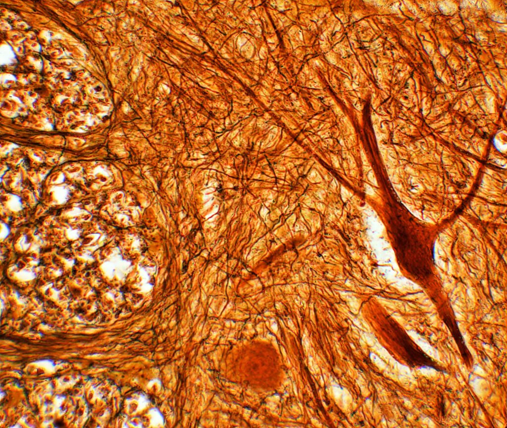
A silver-stained neural tissue section photographed at high magnification, processes branching across the field.
Stained section from a rodent tissue block. Almost every healing claim in this Library was scored by a person looking at an image like this one, which is why blinded assessment matters so much. Photograph: MostlyDross from Springfield, VA, USA / Wikimedia Commons (CC BY 2.0)

04   KPV  /  Alpha-MSH C-terminal tripeptide  /  Mouse (DSS and TNBS colitis)

KPV

Three residues off the end of a hormone, studied almost entirely in chemically injured mouse colon.

What it is

KPV is lysine-proline-valine, the final three residues of alpha-melanocyte-stimulating hormone. The parent hormone has well-described anti-inflammatory activity alongside its pigmentation effects. The published rationale for studying the tripeptide is that it appears to retain part of the anti-inflammatory activity without acting on the receptors responsible for pigmentation.

Reported mechanisms in the primary papers centre on suppression of NF-kappaB signalling in intestinal epithelial cells and macrophages, with downstream reductions in pro-inflammatory cytokine expression.

What the animal literature reports

Nearly all of the in vivo work is murine colitis. The two standard models are dextran sulfate sodium colitis, in which the animal drinks a chemical that damages the epithelial barrier on a schedule the experimenter controls, and trinitrobenzene sulfonic acid colitis, which is hapten-driven and more immune-mediated. Reported outcomes are less body-weight loss in the treated animals, lower composite histological colitis scores, less colon shortening, and reduced cytokine transcript levels in colon tissue.

A substantial fraction of the more recent papers are really delivery papers: the peptide loaded into nanoparticles or hydrogels, with the formulation rather than the peptide as the novel element. Cell culture work in intestinal epithelial lines and in macrophage lines reports the pathway effects described above.

Where the evidence thins

A DSS mouse is a model of chemically induced barrier injury with a defined start date. Inflammatory bowel disease in a person is a chronic relapsing condition of unknown onset with a genetic and microbial background the model does not reproduce. The model is useful for asking whether something reduces acute mucosal inflammation. It is not a small version of the human disease, and results from it should not be read as though it were.

The primary endpoint in most of these papers is a composite histological score assigned by a human observer. Where blinding of that observer is not stated, the effect size should be discounted, and it frequently is not stated. Group sizes are small.

Because formulation differs so widely between papers, cross-study comparison is weak even within the model. There are no published randomized controlled human trials of this tripeptide for any indication.

Evidence snapshot: KPV

Model organisms Mouse, chemically induced colitis; intestinal epithelial and macrophage cell lines
Study scale Small; group sizes commonly six to ten per arm
Human trial status No published randomized controlled trials
Replication status Direction of effect repeated across groups; formulations rarely comparable

Further reading on this page: What a DSS colitis mouse is actually a model of

The question is never whether a compound did something to an animal. Something almost always happens. The question is whether anyone outside the room has ever seen it happen twice.

Nadia Bergeron, Editor

05   LL-37  /  Cathelicidin host-defense peptide  /  Mouse infection and wound models

LL-37

The human cathelicidin, and the reason mouse knockout data cannot speak for it directly.

What it is

LL-37 is the only cathelicidin-derived antimicrobial peptide in humans, a thirty-seven residue amphipathic chain released by proteolytic cleavage from a precursor protein. It is a genuine component of innate immunity, produced by neutrophils and by epithelial surfaces.

It has two distinguishable activities in the literature. The first is direct: it disrupts bacterial membranes. The second is immunomodulatory: it is chemotactic for several immune cell types and alters cytokine responses, including responses to bacterial products. The second is probably the more important one in an intact animal, and it is the harder one to study cleanly.

What the animal literature reports

Mouse models dominate: skin infection, pulmonary challenge with Gram-negative organisms, and systemic infection models, alongside excisional wound healing models reporting faster closure and altered inflammatory infiltrate.

The most informative line of animal evidence is genetic rather than pharmacological. Mice lacking their own cathelicidin gene are more susceptible to certain bacterial infections than wild-type littermates. That is a cleaner form of evidence than administering a peptide, because it asks what happens when the endogenous system is absent rather than what happens when a large exogenous dose is applied.

Cell culture reports broad antibacterial activity against Gram-positive and Gram-negative organisms and activity against some enveloped viruses and fungi.

Where the evidence thins

The in vitro killing concentrations are the central problem. Activity is substantially suppressed by physiological salt concentrations and by serum, so the numbers generated in a low-salt buffer do not carry over to an animal, let alone to a person. At the higher concentrations where killing is reliable, the peptide is also cytotoxic to mammalian cells, which narrows any therapeutic window considerably.

The mouse cathelicidin is not LL-37. It is a different peptide with different properties, which means the knockout work speaks to the class of molecule and to the endogenous system, not to the human peptide as an administered agent. Secondary sources routinely elide this.

Human clinical work is limited to small early-phase studies in specific wound and ulcer contexts. It is not evidence of general antimicrobial benefit, and the peptide has not been shown in controlled human trials to do anything useful about antibiotic-resistant infection.

Evidence snapshot: LL-37

Model organisms Mouse infection, wound and cathelicidin-knockout models; broad cell culture
Study scale Moderate for a preclinical literature; knockout work well established
Human trial status Small early-phase wound studies only
Replication status Endogenous-role findings robust; administered-peptide findings much less so

Further reading on this page: Host-defense peptides and the antibiotic resistance conversation

06   Thymosin α1  /  Thymic immunomodulatory peptide  /  Mouse infection and tumour models; human PBMC culture

Thymosin α1

The one compound here with a real human trial literature, which is exactly why it is worth reading carefully.

What it is

Thymosin alpha-1 is a twenty-eight residue acetylated peptide originally identified in a fractionated extract of thymic tissue. It is an immunomodulator rather than an immune stimulant in any simple sense: the published mechanism involves toll-like receptor signalling and effects on the maturation and function of T cells and dendritic cells.

A synthetic form has been manufactured for decades and is licensed in a number of countries, principally for chronic hepatitis B and as a vaccine adjuvant. That regulatory history is unusual in this Library and changes what kind of reading the evidence deserves.

What the animal literature reports

Mouse work covers immunosuppression models, including chemotherapy-induced and irradiation-induced immune depletion, models of invasive fungal infection, viral challenge models, and transplantable tumour models. Reported outcomes are shifts in cytokine profile, changes in immune cell populations, and in some studies survival differences.

There is also rodent sepsis work, most often caecal ligation and puncture, reporting altered mortality and inflammatory markers. Cell culture in human peripheral blood mononuclear cells reports effects on dendritic cell maturation and on T cell responses, which is the mechanistic backbone of the clinical rationale.

Where the evidence thins

The human literature is genuinely mixed, and that is the important sentence. Early and mid-size trials produced encouraging signals in several settings, and larger or better-controlled work has repeatedly failed to confirm them at the same magnitude. Critical care in particular is a field where early promising immunomodulator results have a long history of not surviving replication.

Rodent sepsis models are among the weakest predictors of human outcome anywhere in preclinical research. Caecal ligation and puncture in a young inbred mouse is not the physiology of an older person with comorbidities in an intensive care unit, and the field itself has said so repeatedly.

The presence of a licensed product in some jurisdictions does not settle the scientific question, and it should not be read backwards as validation of every preclinical claim. What it means is that this is the one compound here where the honest answer to what the human evidence shows is not none, but contested.

Evidence snapshot: Thymosin α1

Model organisms Mouse infection, tumour and sepsis models; cultured human immune cells
Study scale Substantial preclinical and clinical literature by the standards of this Library
Human trial status Multiple randomized trials exist; results mixed and contested
Replication status Preclinical direction consistent; clinical confirmation inconsistent

Further reading on this page: The Sample Size column: what n equals eight buys you, and what it does not

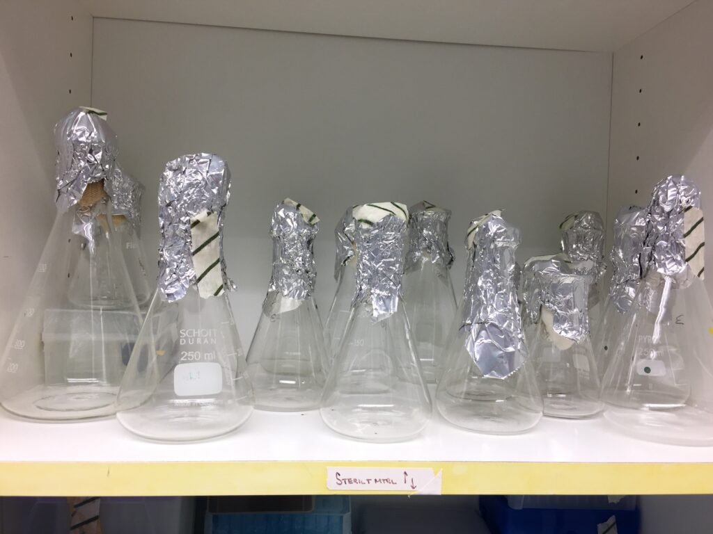
Foil-capped conical flasks cooling on a rack after autoclaving.
Sterile glassware after autoclaving. Cell culture is where most of these compounds were first characterised, and where the concentrations bear no fixed relationship to anything in an animal. Photograph: Pilarbini / Wikimedia Commons (CC BY-SA 4.0)

07   MOTS-c  /  Mitochondrial-derived peptide  /  Mouse metabolic models; cultured myotubes

MOTS-c

A peptide encoded in mitochondrial DNA, studied for about a decade, mostly in mice on high-fat diets.

What it is

MOTS-c is a short peptide of sixteen residues encoded within the mitochondrial 12S ribosomal RNA region. It belongs to a small family of mitochondrial-derived peptides described since the early 2000s, which is to say the entire field is young by the standards of biochemistry.

The published account of its behaviour is that under metabolic stress it moves to the nucleus and participates in transcriptional regulation, with effects converging on AMP-activated protein kinase signalling and folate-methionine metabolism. That is a mechanistically interesting claim and it is still being worked out in the primary literature.

What the animal literature reports

The core in vivo work is mouse metabolic. High-fat-diet models report reduced weight gain and improved glucose tolerance in treated animals. Ageing mouse studies report improvements in physical performance measures such as treadmill capacity and grip strength. Tissue-level work reports transcriptional changes in skeletal muscle and adipose tissue consistent with the proposed mechanism.

Cell culture is largely cultured myotubes and hepatocytes, reporting changes in glucose uptake and in the metabolic pathways named above. Human work exists but is observational: circulating concentrations have been measured across age groups and in various metabolic states and reported to correlate with several outcomes.

Where the evidence thins

This is a young field with a small number of productive groups, several of which share senior authors. That is normal for a new area and it is also exactly the condition under which apparent consensus can form without independent confirmation. Very little of this work has been repeated by laboratories with no connection to the originating group.

The endpoints are metabolic surrogates. Glucose tolerance in a high-fat-diet mouse is a legitimate measure of something; it is not a health outcome, and the diet-induced obese mouse is a model with well-known limitations as a stand-in for human metabolic disease.

The human observational work is the weakest link in the chain as it is usually presented. A peptide whose circulating concentration falls with age and correlates with fitness may be a marker rather than a cause, and no randomized trial of administering it to people has been published.

Evidence snapshot: MOTS-c

Model organisms Mouse, diet-induced obesity and ageing; cultured myotubes and hepatocytes
Study scale Small; a young literature concentrated in few groups
Human trial status None. Human data is observational and correlational
Replication status Limited independent replication to date

Further reading on this page: Mitochondrial-derived peptides: a young field, assessed honestly

08   Humanin  /  Mitochondrial-derived peptide  /  Rat and mouse neuroprotection and metabolic models

Humanin

Most of the striking results belong to a laboratory-made analogue rather than to the peptide itself.

What it is

Humanin is a peptide of twenty-four residues encoded within the mitochondrial 16S ribosomal RNA region. It was first described from tissue that had survived in a brain affected by Alzheimer disease, which is where its name and much of its early framing come from.

An important practical point for anyone reading about it: several engineered analogues exist in the literature, including potency-enhanced variants produced by single-residue substitution. A large share of the most quoted results were obtained with an analogue, not with the native peptide. Results with one are not results with the other, and secondary sources conflate them constantly.

What the animal literature reports

The neuroprotection work is mostly rat and mouse, and mostly central. Reported models include intracerebroventricular administration alongside amyloid-beta challenge, and cerebral ischaemia models, with outcomes reported as reduced neuronal loss on histology and better performance on spatial memory tasks such as the water maze.

There is a separate metabolic literature in mice reporting effects on insulin sensitivity and on glucose handling, and work in rodent models of retinal degeneration reporting preserved photoreceptor layers. Cell culture reports protection of neuronal cell lines against several toxic insults.

Where the evidence thins

Rodent cognitive endpoints are noisy. Water maze performance moves with handling, with time of day, with the strain used, and with who scored the video. Small studies on such endpoints, unblinded, are the easiest place in preclinical research to find an effect that is not there, and this literature contains a good deal of exactly that design.

Route of administration is the other issue. Delivering a peptide directly into the ventricles of a rat brain answers a mechanistic question and says nothing about what peripheral administration would do, because the thing that would have to happen next, crossing into the central nervous system in a meaningful amount, is the part that was bypassed.

Human data is observational. Circulating concentrations decline with age and have been reported to associate with various measures of health. Association is not effect, and no randomized human trial of administration has been published.

Evidence snapshot: Humanin

Model organisms Rat and mouse, central administration; mouse metabolic; neuronal cell lines
Study scale Small, with noisy behavioural endpoints
Human trial status None. Human data is observational and correlational
Replication status Analogue and native peptide results routinely conflated in secondary sources

Further reading on this page: Mitochondrial-derived peptides: a young field, assessed honestly

Wooden card catalogue drawers with brass label holders, viewed at an angle down the aisle.
A card catalogue. Tracing a claim back to the paper that first made it, rather than to the tenth source that repeated it, remains the single most useful thing a reader can do. Photograph: Ted Eytan / Wikimedia Commons (CC BY-SA 2.0)

09   House style

How to read these summaries

Every profile above is built the same way, and the structure carries meaning. The first section describes the molecule without evaluating it. The second reports what has been published in animals and in cultured cells, naming the species and the model each time, because a result in a Sprague-Dawley rat and a result in a cultured human fibroblast are different kinds of claim and should never be summarised in one sentence.

The third section is the one most publications leave out. It states what is missing: which findings have never been repeated outside the originating laboratory, where group sizes were too small to detect the effect being claimed, where the assessor was not blinded, and whether any randomized human trial exists at all. When the answer to that last question is no, the profile says no rather than saying that human research is ongoing, which is a phrase that can be written about anything.

The hedged language is deliberate and it is not evasion. Reported, observed in, has been described, remains unreplicated: each of those phrases marks the difference between what a paper found and what the world is like. Papers report findings. Findings become knowledge when other people, with no stake in the first result, find the same thing.

Absence of evidence gets stated plainly here rather than glossed. For most of these compounds there is no rigorous human evidence, and saying so is not a criticism of the researchers involved. Preclinical work is supposed to generate hypotheses. It becomes misleading only when it is read as though it had already tested them.

Keep reading

The reporting behind these entries

Library profiles are deliberately short. The longer arguments, about replication, study design and publication history, run in Dispatches.

Browse Dispatches